 |
Medical Retina and the Posterior Segment:
Case fifteen
|
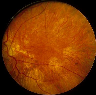
This 20 year-old man was referred by his family doctor because of poor nigh vision. On examination, his visual acuity was 6/9 in both eyes. Electrophysiology showed decreased a and b waves in electroretinography and there was abnormal dark adaptation. The feature of the left fundus is shown above.a. What does the picture show?
There is widespread loss of the retinal pigment epithelium and choriocapillaris with clumps of pigment beneath the retina (in the choroid).
b. What is the most likely diagnosis?Choroideremia.c. What is the differential diagnosis?The condition is an X-linked recessive (it is caused by a deletion in the Xq21 band of the X chromosome) disorder characterized by bilateral progressive chorioretinal degeneration with night blindness, peripheral field loss and loss of central vision in the late stage.
The diagnosis is based on the typical fundal appearance. The
ERG shows decreased a and b wave. Fluorescein angiography shows loss of the choriocapillaris.
The main conditions to be considered are:d. What is the visual prognosis in this patient?
- retinitis pigmentosa (bone spicules pigments are seen in the retina which is absent in choroideremia)
- gyrate atrophy (rare autosomal recessive, high ornithine levels in the body fluid, it is caused by a deficiency of the mitochondrial enzyme ornithine aminotransferase. The fundi show sharply defined area of RPE loss )
- degenerative myopia
- ocular albinism
In choroideremia, the patient becomes symptomatic in the first decade of life with reduced night vision. The visual field shows progressive loss with age. The visual acuity typically remains good until the fifth decade.
| Click here for the questions | Click here for the main page | Click here for FRCOphth/MRCOphth
/FRCS tutorials |