Figure 1 |
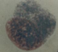
Figure 2 |
| Paediatric ophthalmology: Case Six |
|
Figure 1 |
Figure 2 |
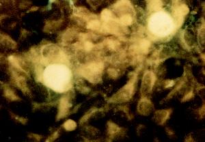
This is the immunofluorescent staining on the conjunctival scrapping of a two-week old baby who presented to the casualty with a discharging right eye.a. What does the picture show and what is the diagnosis?
The immunofluorescent staining shows fluorescent inclusion bodies within the epithelial cells. The diagnosis is neonatal chlamydial conjunctivitis.b. How is the disease acquired?
The inclusion body is also demonstrated in Figure 2 with a Giemsa staining. It appears as a basophilic cytoplasmic inclusion.Neonatal chlamydial conjunctivitis is caused by infection with Chlamydial trachomatis (serotypes D - K). The baby is infected during passage through the birth canal.
It typically presents as a mucopurulent papillary conjunctivitis. The incubation is usually 5 days but may vary from 4 to 12 days.
c. What other diseases may be caused by this infection?Pneumonia and otitis media
d. How would you manage this baby?Management should include the treatment of the conjunctivitis as well as the prevention of pneumonia. Therefore, topical treatment alone is not sufficient. The recommended treatment is with oral erythromycin (50mg/kg/day) for two weeks.
The parents should also be treated with oral erythromycin or tetracycline. Tetracycline should be avoided in breast-feeding mother.
Click here for the questions Click here for the main page Click here for FRCOphth/MRCOphth
/FRCS tutorials