 |
Medical Retina & the Posterior Segment:
Case eighteen
|
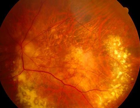
This 35 year-old woman presented to the casualty with a one-week history of painful right eye and decreased vision. She was found to have a left iritis and the above fundal appearance.a. What does the picture show?
There is extensive vasculitis with exudate giving the "candle-wax dripping" appearance.b. What is the most diagnosis?Retinal vasculitis caused by sarcoidosis.Sarcoidosis is a granulomatous, systemic disease of unknown cause. Ocular involvement occurs in 35% of patients and of these one quarter would have posterior segment involvement. The posterior segment lesions include retinal vasculitis with typical candle-wax dripping, posterior uveitis, chorioretinitis, choroidal granuloma and papillitis.
c. What non-invasive investigations would you perform to support your diagnosis?The two non-invasive tests that can support the diagnosis are:
- Serum angiotensive converting enzyme (ACE) level. ACE is produced by the granuloma and is elevated in 90% of patients with sarcoidosis. However, it is not specific to sarcoidosis and can be elevated in other granulomatous diseases
- Chest X-ray. It is abnormal in 80% of patients with bilateral hilar lymphadenopathy, interstitial infiltration and fibrosis.
d. How can the above appearance affect the patient's vision?Retinal vasculitis can affect the vision through:
- macular ischaemia and oedema
- occlusion of the retinal vessels
- peripheral retinal ischaemia leading to neovascularization and vitreous haemorrhage
| Click here for the questions | Click here for the main page | Click here for FRCOphth/MRCOphth
/FRCS tutorials |