Figure 1 |
Medical retina and posterior segment: Case nine
|
Figure 1 |
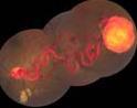
Figure 2 |
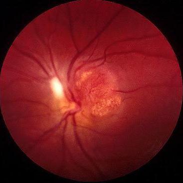
This 30 year old woman was referred by her optician because of this left optic disc appearance. She was otherwise fit and well. Her visual acuity was 6/6 in the right eye and 6/12 in the left. Her mother died of brain haemorrhage at the age of 40 and a brother had an operation for renal tumour.a. What does the picture show and what is the most likely diagnosis?
There is a round reddish tumour arising from the optic disc (Figure 1). The appearance is typical of a capillary haemangioma of the optic disc. The history suggests that this is a von Hippel-Lindau's angioma.Capillary angioma outside the optic disc usually presents as an orange red tumour fed by a dilated retinal artery and drained by an engorged retinal vein (Figure 2)
b. How would you investigate this patient?von Hippel-Lindau's syndrome is a type of phakomatosis associated with multiple organ haemangblastoma (including retina, cerebellum, kidney), cysts (in the pancreas, kidney, lung and liver), phaeochromocytoma and renal cell carcinoma.The investigation show aim to look for potentially life-threatening conditions and this include:
- MRI scan for cerebellar haemangioma, phaeochromocytoma and renal cell carcinoma
- 24 hours urine VMA for phaeochromocytoma
c. How is this condition inherited?Autosomal dominant condition with incomplete penetrance. The defect is thought to reside on the short arm of chromosome 3.
d. What is the visual prognosis of this condition?Visual loss and distortion can occur due to formation of exudates in the macula or leakage of fluid producing serous retinal detachment. Treatment of optic disc capillary haemangioma is difficult as the use of photocoagulation runs the risk of damaging the optic nerve.
Click here for the questions Click here for the main page Click here for FRCOphth/MRCOphth
/FRCS tutorials