Figure 1 |
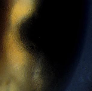
Figure 2 |
Eyelids & the Anterior Segment:
Case twenty six
|
Figure 1 |
Figure 2 |
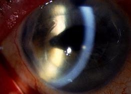
This 65 year-old woman complained of deteriorating vision in her left eye (Figure 1). She had had an operation for cataract one year earlier. Figure 2 is the appearance of the cornea at higher magnification.a. What is the diagnosis?
Pseudophakic bullous keratopathy.b. What conditions would increase the development of this condition?Figure 1 shows the presence of an anterior chamber lens and hazy cornea. Figure 2 shows cystic changes on the corneal surface caused by corneal oedema. These features are the results of endothelial decompensation. The two main complaints in this condition are visual loss and pain.
Pseudophakic bullous keratopathy is increased in:
- Endothelial damage resulting from complicated intraocular injury (as in this case). The presence of anterior chamber lens also increases the rate of endothelial loss.
- Pre-existing endothelial dystrophy such as Fuch's endothelial dystrophy.
- Severe and chronic intraocular inflammation
- Endophthalmitis
c. How would you manage this condition?Management may include any of the following:
- Observation. If the patient suffers no pain and is able to function with one good eye and does not wish to have surgical treatment.
- Medical. Swelling of the cornea may be treated with hypertonic sodium chloride (5%) which is useful at the early stage. Pain caused by ruptured bullae can be treated with lubricants or bandage contact lens.
- Surgery. Penetrating keratoplasty is the definitive treatment for this condition. The anterior chamber lens is also exchanged to prevent it causing damage to the graft endothelium. The new lens may be sulcus fixated if there is enough capsular support or fixated to the iris or sclera.
| Click here for the questions | Click here for the main page | Click here for FRCOphth/MRCOphth
/FRCS tutorials |