 |
Neuro-ophthalmology: Case twelve
|
f
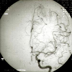
This 32 year-old woman had had two years history of recurrent left sided headache preceded by photopsia. She was put on prophylactic medication for classical migraine without effect. A cerebral angiography was performed after she experienced a transient right hemiplegia. a. What does the cerebral angiography show?
Arteriovenous malformation in the left parietal lobe.Arteriovenous malformations (AVMs) are developmental abnormalities of blood vessels. About 85% of AVMs are located superficially in the cerebral hemisphere. The rest are deep in the cerebral hemisphere or in the posterior fossa.
b. How does this condition present clinically?AVMs usually come to medical attention because of one or a combination of the following problems:Headache usually of migrainous type can occur in 10% of AVMs and is thought to caused by recurrent small bleeding. They characteristically begin during the second decade of life. When the visual pathway is involved photopsia is common.
- intracranial haemorrhage
- seizures
- focal neurological deficits
- headache
c. How could it be treated?Depending on the location of the lesion, it can either be surgically excised, embolized or treated with radiotherapy. In this patient, excision of the lesion would have led to visual defect and high cortical dysfunction and therefore she was treated with radiotherapy.Radiotherapy is thought to work by causing reactive intimal proliferation leading to obliteration of the vessel lumen.
| Click here for the questions | Click here for the main page | Click here for MRCOphth/FRCS tutorials |