 |
| Paediatric Ophthalmology: |
| Case eighteen |
|
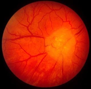
This 10 year-old boy was referred by the neurologist for a retinal examination. He was on treatment for epilepsy. a. What does the fundal show?
Retinal astrocytoma.
The picture shows a white lesion on the retinal nerve fibre layer. This is caused by the hamartomatous proliferation of astroglia.
b. What is the most likely diagnosis of the underlying condition?Tuberous sclerosis (Bourneville's syndrome).c. How is this condition inherited?
The presence of retinal astrocytoma in a patient with epilepsy strongly suggests tuberous sclerosis. The classic triad of tuberous sclerosis are: epilepsy, mental retardation and sebaceous adenoma of the face.Tuberous sclerosis may be inherited in an autosomal dominant fashion with variable penetrance or arises as spontaneous mutation.Autosomal dominant tuberous sclerosis are of two types depending on the abnormal chromosome:
- type 1 : chromosome 9q33-34
- type 2: chromosome 11q23
d. What other ocular condition may occur?Apart from retinal astrocytoma, the following may be seen:
- eyelid angiofibroma
- sector iris hypopigmentation
- retinal pigment epithelium defects
e. List some of the systemic diseases seen with this condition?Skin lesion:Central nervous system:
- sebaceous adenoma (these are angiofibroma composed of fibrous tissue and capillary channels)
- ash-leaf spots (hypopigmentation of the skin best seen with ultraviolet or Wood's light)
- shagreen patches (fibrous thickening)
- periungual fibroma
Renal:
- mental retardation
- epilepsy
- central nervous system hamartoma
Cardiac system:
- cysts
- tumours
- rhabdomyoma
| Click here for the questions | Click here for the main page | Click here forMRCophth / MRCS tutorials |