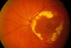
| This 75 year-old man presented to the eye casualty with a one week
history of distorted left vision. His visual acuity was 6/12 in the left
eye with the above posterior segment appearance. He had no history of hypertension
or diabetes mellitus.
a. What is the most likely diagnosis?
Age-related macular degeneration with subretinal neovascular
membrane.
The above picture shows circinate exudate in the macula with a greyish
lesion in the centre representing the choroidal neovascular membrane with
or without pigment epithelial detachment.
b. What other condition may be responsible for the above appearance?
Circinate exudate in the macula can be caused by two mechanisms:
-
Breakdown of the inner blood retinal barrier (tight junctions between the
retinal vascular endothelial cells). For example: diabetes mellitus, retinal
vein occlusion, chronic uveitis and radiation retinopathy.
-
Breakdown of the outer blood retinal barrier (tight junctions between the
adjacent retinal pigment epithelial cells). For example: choroidal neovascular
membrane due to macular degeneration, choroidal rupture, choroidal melanoma
and presumed histoplasmosis syndrome.
c. How would you manage this condition?
The patient should be assessed for her suitability for laser
treatment (which is the only statistically proven effective treatment).
Fluorescein angiography should be requested to look for the type and
location of the subretinal neovascular membrane. Patient who will benefit
from laser will be those with classical choroidal neovascularization which
is located outside the fovea. Those with occult choroidal neovascular membrane
(in which the membrane is diffuse and poorly defined) or subfoveal lesion
are not suitable for laser.
In centre where indocyanine green (ICG) videoangiography is available,
those with occult choroidal neovascular membrane as seen on FFA should
undergo ICG. ICG gives a better visualization through the pigment and blood
and may localize area of choroidal neovascularization for laser therapy.
The visual prognosis is usually poor even in those who can be treated
with laser as the choroidal neovascularization has a high recurrence rate
(about 50% within 3 years)
|