Paediatric ophthalmology: Case nine


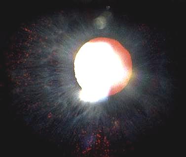
This is the slit-lamp appearance of a ten year-old girl whose brother had bilateral poor vision and nystagmus.

a. What does the picture show?

The iris shows diffuse loss of pigment giving a moth eaten appearance.
b. What is the diagnosis?
The patient is a carrier of X-linked ocular albinism.

In X-linked ocular albinism, the affected male has iris transillumination, nystagmus and hypopigmented fundus. Macular hypoplasia is a common cause of decreased vision. Strabismus and decreased binocularity (due to excess decussation at the chiasm) are also common.

Carrier female demonstrates partial iris transillumination and retinal pigment epithelium defects. Otherwise, the vision is normal.


c. What other forms of this condition do you know of?

Albinism can be divided into:
  • ocular albinism (affecting the eye only and is often inherited as X-linked or autosomal recessive)
  • oculocutaneous albinism (systemic involvement with white hair and skin and is usually inherited in autosomal recessive fashion)
  • Clinically, albinism can be divided into:
  • tyrosinase-positive (in which the there is progressive increase in pigmentation as the body is able to produce melanin, the visual acuity may show improvement due to increased pigmentation in the retinal pigment epithelium)
  • tyrosinase-negative (in which the body is incapable of producing melanin and the visual acuity is very poor
  • Click here for the questions Click here for the main page Click here for MRCOphth / MRCS tutorials