Xanthelasma occurs commonly in the periorbital region and therefore
the pathologist is never short of slides for this condition.
The features to look for are:
-
multiple clear cells in the dermis (typically
around a blood vessel); these cells are also called foam
cells and they are macrophages which are lipid filled. Due to the
prepartion of the tissue, the lipid component of the cells are removed
by the alcohol and therefore the cells appears empty.
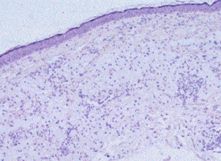
Low magnification H&E
Xanthelasma showing cells in the dermis. At higher
magnification, the cells can be seen as lipid-laden
macrophages. |
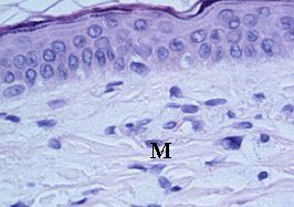 .... ....
High magnification H & E
Presence of lipid-laden macrophages (M) which appear
to
have clear cystoplasm due to removal of lilpid during
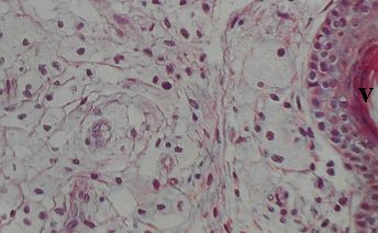
tissue preparation. The right picture shows the presence
of multiple foam cells next to a blood vessel (V). |
Common questions in the viva:
-
How would you manage a patient with xanthelasma? (blood tests
for cholesterol, excision of lesion or laser or chemical treatment)
|