Molluscum contagiosum is a popular topic because the lesion
has very characteristic appearance. The opening question may be: " This
is the lesion taken from a patient with recurrent (follicular) conjunctivitis.
What can you see?"
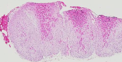
The features to look for are:
-
epidermis acanthosis (hyperplasia) which grows
into the dermis to form multiple lobule
-
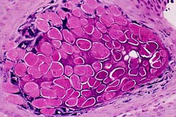
intracytoplasmic inclusion bodies which are
eosinophilic (stianed pink with H & E) at the base but becomes more
basophilic (stained blue with H & E) at the superficial layer
-
central crater into which the inclusion bodies discharge their content