Fungal keratitis is uncommon in the UK. However, because the
corneal biopsy or button of fungal keratitis show characteristic hyphae
with silver stains; such slide had appeared in the examination.
Two conditions are responsible for most of the fungal keratitis:
-
corneal trauma with contaminated vegetation (Fusarium and Aspergillus
are the commonest)
-
pre-existing corneal diseases such as severe dry eyes or patients on chronic
steroid use (Candidas is usually involved)
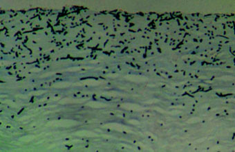
Corneal infected with Fusarium showing hyphae. The cornea
is
also infiltrated by neutrophils. The specimen is stained
with
Grocott hexamine (methenamine) silver.
|
Common viva questions:
-
When would you suspect fungal keratitis? (History and clinical signs.)
-
A man with poorly controlled diabetes mellitus developed orbital cellulitis
which was not responsive to antibiotic. A MRI scan revealed opacity in
the ipsilateral maxillary sinus with bony destruction. What is the differential
diagnosis? (You will be expected to mention mucormycosis.)
-
An intravenous drug abuser complained of blurred vision. Examination revealed
an unilateral dense vitritis. What is the differential diagnosis? (Include
Candidas as a cause for endogenous enophthalmitis in your answer.)
|