a
Fuchs endothelial dystrophy
q
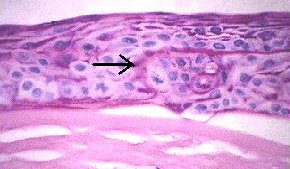
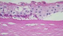
This is the most common endothelial dystrophy seen in clinical practice.

In the examination, you are likely to be asked about the histological differences between Fuchs endothelial dystrophy and pseudophakic bullous keratopathy. As both conditions will require full thickness keratoplasty in advanced stages, you are likely to be given slides showing the following signs:

  • bullae in the epithelium
  • intraepithelial basement growth from chronic epithelial oedema
  • absence or very reduced endothelium


Bullous keratopathy. Arrow indicates intraepithelial basement membrane. 
This is also seen in conditions such as 
finger-dot dystrophy. (PAS stain)

Bullous keratopathy. The epithelium 
shows multiple cystic spaces. Unlike the epithelium, spaces in stroma are common artifact and do not imply stromal oedema. (PAS stain)

The two conditions differ in that Fuchs endothelial dystrophy has the following additional signs:
  • guttata in the endothelial layer
  • thickened Descemet's membrane.


Fuchs endothelial dystrophy showing thickened 
Descemet's membrane, guttata and absent 
endothelium. (PAS)

High magnification showing guttata and 
thickened Descemet's membrane. 

Common viva questions:
  • What other types of endothelial dystrophy do you know of?
  • How do you differentitate Fuchs dystrophy from other causes of endothelial dystrophy such as pseudophakic bullous keratopathy?
Return to the cornea index