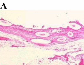
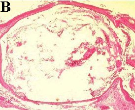
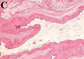
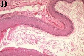
a
Epidermoid and dermoid cysts
q
    Epidermoid and dermoid cysts are common surgical specimens sent for histopathological analysis.
       
      • Dermoid cysts are usually congenital (a type of choristoma ie. lesion containing  mature tissue but found at an abnormal location). They may be found in the eyelid, peripheral cornea or orbit. In the latter, the most common location is in the superotemporal quadrant. The content of the wall comes from desquamation of the epithelium and secretion of the sebaceous glands lining the wall. 
      a
      • Epidermoid cysts come from squamous epithelial cells that have penetrated deep into the 

      • dermis  for example after skin surgery, trauma or congenital. The content of the cyst is mainly keratin as only the cysts do not contain dermal structures such as hair follicle or sebaceous glands.
      a
    Histologically, the two differs in the structures found in the lining of the wall. 

    Features common to both are:

       
      • keratinized and stratified squamous epithelium
      • eosinophilic (pinkish when stained with H&E) content within the wall
      n
    However, in dermoid but not in epidermoid the following are present:
       
      • presence of sebaceous glands, sweat glands and hair follicles ie. structures from

      • the dermis.

     
    Test yourself pictures
    ( Using the information above decide whether the slide is an epidermoid or a dermoid cyst?)

    a
    Additional features which may be present:

    • inflammatory cells surrounding the cysts (in both) if there is rupture of the cyst or the content of the cyst has leaked out. Foreign body giant cells may be seen.
Return to the main page