H&E and PAS are the two most commonly used stains
in histopathology.
Haematoxylin and eosin (H & E)
This is the most commonly used stain in routine pathology. Haematoxylin,
a basic dye stains acidic structures a purplish blue. Nuclei (DNA), ribosomes
and rough endoplasmic reticulum (with their RNA) are therefore stained
blue with H&E. Eosin, in contrast is an acidic dye which stains basic
structures red or pink. Most cytoplasmic proteins are basic and therefore
stained pink or pinkish red. In summary, H&E stains nuclei blue and
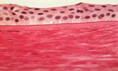
cytoplasm pink or red. The following specimen shows a normal skin stained
with H&E.
PAS (periodic acid-Schiff)
This stain is versatile and has been used to stain many structures including
glycogen, mucin, mucoprotein, glycoprotein, as well as fungi. PAS is useful
for outlining tissue structures--basement membranes, capsules, blood vessels,
etc. As it stains many structure; this can give rise to a high background.
It is very sensitive, but specificity depends upon interpretation. The
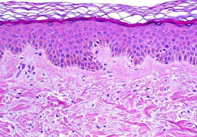
following specimen is a normal cornea that has been stained with PAS and
the basement membrane of the epithelium is highlighted.