Acanthamoeba keratitis is the most serious infective keratitis associated
with contact lens wear. Although it is uncommon (Pseudomonas aeruginosa
is the most common organism involved in contact lens associated infection
followed by other bacteria), it is difficult to treat and may be mis-diagnosed
as herpertic keratitis at the early stage. The examiner will expect the
candidates to know this organism well as well as the associated clinical
signs.
In the examination, you may be given slide showing cornea infected with
acanthamoeba or simply ask to discuss the management of a patient with
contact lens related corneal infection.
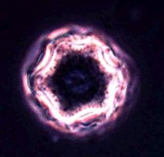
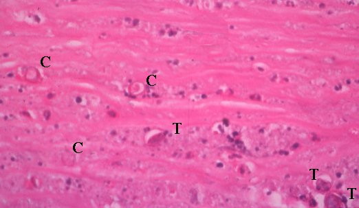
cyst |
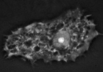
trophozoite |
Acanthamoeba exists in two forms: cyst (which is double-walled) and
trophozoite. Both may be seen in corneal scrap or biopsy.
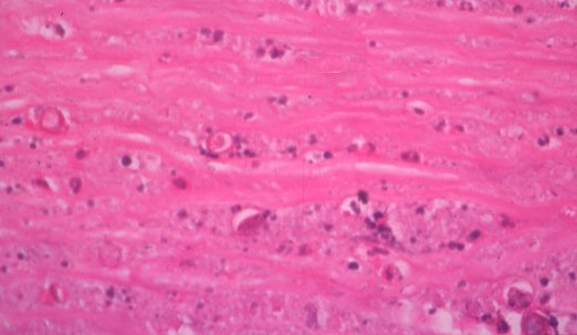
A specimen of cornea showing acanthamoeba cysts and trophozoite.
Click on the picture to see if you have found the organisms. |
Common viva questions:
-
How can you identify acanthamoeba in cornea? (Using special
stains, see the section on stains
for microbes; culture using nutrient poor agar overlay with E.coli
and immunofluorescent staining on the corneal scrap. Confocal microscopy
may be used to identify acanthamoeba in cornea..)
-
What factors and signs may alert you to the possibility of acanthamoeba
keratitis?
-
What are the treatment options for acanthamoeba keratitis?
|