Opticociliary shunt (collaterals) vessels
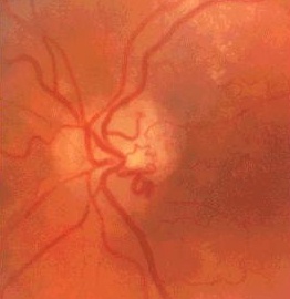 |
The disc shows dilated venous loop.
Unlike neovascularization,
there are no thin or wispy.
Look for:
old central retinal vein occlusion such flamed
haemorrhages, tortous retinal vein or
panphotocoagulation scars for ischaemiccentral
retinal vein occlusion
evidence of retrobulbar tumour such as proptosis and choroidal
foldings.
|
Questions:
1. How can you differentiate shunt vessels from neovascularization?Answer
2. What is the differential diagnosis of opticociliary
shunt vessels?