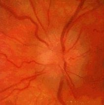
The optic disc margin is blurred. There is hyperaemia
with or without splinter haemorrhage(s). The venous pulsation is absent.
Note:
it is safer to state that the patient has optic swelling rather than
papilloedema unless you have evidence of raised intracranial pressure.
Sometimes you may encounter patients with indistinct
optic disc without disc swelling or haemorrhages. In such cases,
consider pseudo-papilloedema as in patients with high hypermetropia.
Look for:
hypertensive changes in malignant hypertension
extensive haemorrhages in central retinal vein occlusion
examine the contralateral eye for similar changes. The presence
of bilateral disc changes strongly suggests intracranial hypertension as
the cause. The most common case seen in the examination is a young overweight
woman with benign intracranial hypertension.
optic atrophy in the contralateral eye ie. Foster-Kennedy's
syndrome which is classically caused by meningioma of the optic canal (in
the eye with optic atrophy). However, in both examination and real life
the most common cause is sequential ischaemic optic neuropathy.
Questions:
1. What are the causes of a indistinct optic disc margin?Answer