High myopia
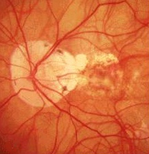 |
The optic disc has irregular peripapillary
atrophic crescent (this is due
to failure of the retinal pigment epithelium and choroid to reach the disc margin). There are thin white lines (termed lacquer crack caused by breaks in Bruch's membrane) extending from the optic disc. Other signs to look for:
neovascularization and haemorrhage) the cavity (peripheral chorioretinal scars or indentation) |
Questions:
1. What is the definition of high myopia?Answer
2. What are the causes of myopia?