Spastic paresis
.
The tone is increased in both legs. The lower limb muscles
are weak. There are hyper-reflexia with upgoing plantar (positive Babinski's
reflex). The sensation is impaired (examine for a sensory level). On attempted
walking, there is scissoring of the gait (there is crossing over of the
feet with dragging of the toes).
Other examination:
-
There are many causes of spastic paresis, the most important
being spinal cord compression either from tumour or trauma. However, in
the MRCOphth, most would be caused by multiple sclerosis. Therefore, mention
you like to to examine the eyes for optic atrophy, internuclear ophthalmoplegia
or nystagmus
-
mention you like to examine the back for any scar. The patient
may have neurofibroma of the spine removed and there may be other stigmata
of neurofibromatosis.
. |
Hemiplegia
.
The affected limb may have abnormal posture with external
rotation and extension. The tone of the affected leg is increased. The
muscle power is weak. There is hyper-reflexia with upgoing plantar response
on the same side. The sensation may be abnormal if the sensory cortex is
also involved. On walking the foot tends to circumduct and rotate in a
semi-circle with each step.
Other relevant signs:
-
hemiplegia of the ipsilateral upper limb
-
ipsilateral upper seventh nerve palsy
-
hemianopia
-
mention you like to examine the cardiovascular system especially
for pulse, heart sounds and carotid artery for possible source of embolism.
. |
Combined
upper and lower motor neurone lesion
.
The patient has absent knee and ankle jerks. The plantar
responses are upgoing. The sensation may be normal or impaired depending
on the cause.
When you elicit these signs, consider the following possibilities:
-
diabetes mellitus. This is by far the most common and the
signs are caused by peripheral neuropathy and cerebrovascular accident
both of which are common in diabetes mellitus. The affected side will have
hemiplegia. The sensation is impaired with possible stocking distribution.
In severe sensory loss, there may be Charcot's joint of the ankle (due
to repeated painless trauma) and foot ulcer
-
Fridriech's ataxia
-
subacute combined degeneration of the spinal cord. This uncommon
condition is due to vitamin B12 deficiency. The muscles may be spastic
and there may be optic atrophy.
-
motor neurone disease
-
tabes dorsalis
. |
Friedriech's ataxia
.
The candidates are usually asked to either examine
the lower limbs or test the cerebellar function.
The patient has pes cavus (high arches of the feet). There
are weakness of the lower legs. The knee and ankle jerks are absent but
the plantar is upgoing. There is impaired sensation to vibration and joint
sense. The gait is wide-spaced due to ataxia.
Other signs:
-
cerebellar signs are prominent with nystagmus, scanning speech,
intention tremor and past-pointing
-
mention that you would like to look for optic atrophy
-
other associated features: diabetes mellitus is common and
the patient may have diabetic eye disease and cardiac diseases
. |
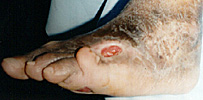
Diabetic foot
.
This is usually an extension of the fundal examination
in a patient with diabetic retinopathy. The candidate is usually asked
which part of the body he would like to examine to look for diabetes-related
complications.
The patient has ulcer over the pressure point of the foot.
They may be missing toe due to amputation. The ulcer may be ischaemic or
neurotrophic:
-
in ischaemic ulcer, the skin is cold with absent pedal dorsalis
and tibilias posterior.
-
in neurotrophic ulcer, there are sensory loss to pain, temperature
and light touch typically in a stocking distribution.
Look for:
-
disorganized ankle joint (Charcot's joint) form joint position
and vibratory sensory loss.
|
|