When presented with an image; the following steps are recommended
as in the interpretation of CT scans:
-
What type of image is it? MRI scans (T1 or T2 weighted)
-
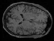
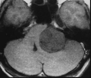
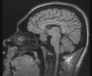
Which plane the image is in? axial, sagittal or coronal (see
pictures below)
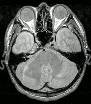
Axial scan |
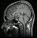
Saggital scan |
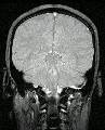
Coronal scan |
-
Describe the image: In ophthalmology, most of the pathology will
be focused on the globe, orbit or the brain. If it were the viva,
ask the examiner for some histories.
-
Examine the following strucutres in order and always compare the
two sides for asymmetry(ies):
-
globe
-
extraocular muscle enlargement
-
space occupying lesion in the orbit
-
bony lesion
-
brain lesion
Click the following pictures for Questions
and Answers.
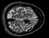
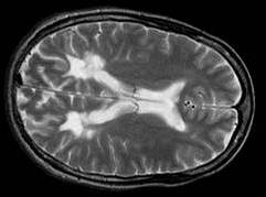
(The experience from past candidates inidcates that the
examiners expect you to be able to differentiate T1 from T2 weighted images.
The most commonly encountered scan in the examination is periventricular
plaques.)
 |
 |
 |
g
a |