( The following are answers to mock examination 4. To return to each question click on the number)
1.
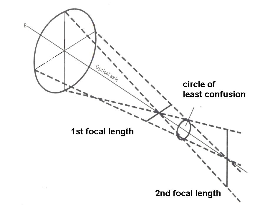
i) Location of the first local line (formed by the meridian with +3.00D) Using the formula 1/u +1/v = 1/fii) Location of the second local line (formed by the meridian with +2.00D) Using the formula in i)b. Location of the circle of least confusion = (length of first focal length + length of second focal length) / 2 = (50 + 100) / 2 = 75 cm behind the lens c. Diameter of the circle of least confusion = (D1 - D2) / (D1 + D2) X aperture of the lens D1 = the lens power that produces the first focal length = 3.00 D D2 = the lens power that produces the second focal length = 2.00 D aperture of the lens = 60 mm = 1/5 (60)
a. The resultant prism is found by adding the powers of the two prisms
b. The resultant prism is found by subtracting the weaker prism from
the
c. The resultant prism is found by adding the powers of the two prisms
d. The resultant prism is found by subtracting the weaker prism from
the
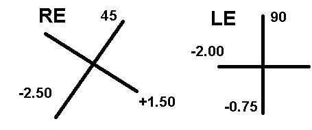
( For horizontal prismatic effects: b. when prisms are placed with their bases in the opposite direction and of3. a. The power crosses translate to spherocylindrical corrections (with minus cylinder) of RE +1.50 / - 4.00 X 135 LE -0.75 / -1.25 X 90 As the working distance is 2/3 metre, -1.50
D is subtracted from the above giving
b. The power crosses translate to spherocylindrical corrections (with
positive cylinder) of
As the working distance is now 1/2 metre,
-2.00D is subtracted from the above
a.
ii. Bigger. In a myope, the
eye has more plus power and using the formula of
b.
ii. When a 15 D lens is used. The retina
magnification = 60/15 = 4X
a. Sturge-Weber's syndrome ( The skull X-ray shows the typical
curvilinear calcification
b. Right retinoblastoma ( There is calcification in the right
posterior pole. In a child with poor
c. Foreign body in the left orbital region ( There is a small
opacity in the left orbit, this may
a. Fluorescence has an absorption peak from 465 to 490 nm (blue region
of the visible spectrum)
b. Venous phase.
The picture shows an area of hyperfluorescence in the macula. The hyperfluorescence assume a smoke stack appearance. This is a classical appearance of central serous retinopathy a. Fully accommodative esotropia = refractive accommodative esotropia
b. Non-refractive accommodative esotropia = accommodative esotropia
with convergence
c. Right lateral rectus palsy
8. a. Hess chart relies on two laws: Sherrington's law ( law of reciprocal innervation)
= the contraction of a muscle is accompanied
Hering's law (law of equal innervation) = innervation
to the extraocular muscle is equal in both
b. Right third nerve palsy ( The field of the affected eye is the small one) c. Right gaze ( The hess chart shows right lateral rectus underaction
and the most likely cause if abducent
|
| Click here for mock examination 4 |
| Click here to return to OSE |