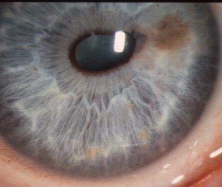
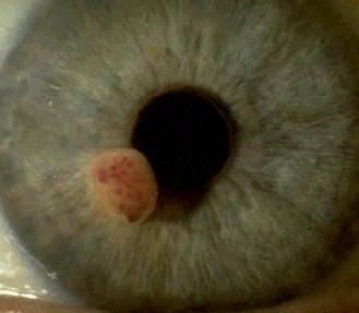
Pigmented lesion of the iris
|
||
|
There is a pigmented lesion on the iris (describe the colour and the edge) which is flat/elevated. Look for signs of malignancy such as abnormal iris vessels around or within the tumour, ectopia uvea and sectorial cataract at the site of the lesion. Note: ectropia uvea may occur in iris lesion which are benign. Further examination:
is at the angle) may be an extension from a lesion behind the iris lesion at the ciliary body with anterior extension. |
Questions:
1.
What is the differential diagnosis of pigmented iris lesion(s)?