Figure 1 |
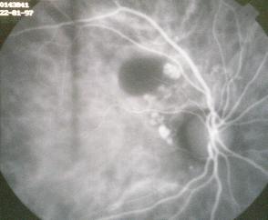
Figure 2 |
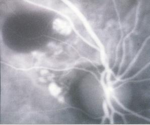
Figure 3 |
This 63 year old Afro-Carribean woman presented to
the eye casualty with a sudden onset loss of left vision. Her visual acuity
was 6/9 in the right eye and HM in the left eye with glasses. Her past
medical history apart from medically controlled hypertension was unremarkable.
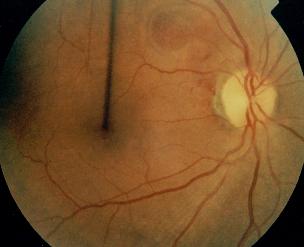
Fundoscopy of the left eye revealed vitreous haemorrhage. In her right
eye, a raised yellowish lesion is seen superotemporal to the optic disc
(Figure 1). An indocyanine green (ICGA) angiography was performed was performed
(Figure 2).
a. What does the ICGA reveal?
The ICGA shows:
- an area of hypofluorescence corresponding to the raised yellowish lesion, this is caused by serous pigment epithelium detachment
- multiple polypoidal structures at the edge of a fine vascular network superotemporal to the optic disc
b. What is the diagnosis?
The history and the ICGA appearance is classical of idiopathic polypoidal choroidal vasculopathy (IPCV).
c. What is the natural history of this condition?
IPCV typically occurs in women who are in their fifth to seventh decade of life and appears to be commoner in black. It is characterized by aneurysmal polypoidal subretinal lesion that arise from the choroidal vessels. Symptoms occur from serosanginuous detachments of the retinal pigment epithelium or breakthrough vitreous haemorrhage.The condition may resolves spontaneously but focal laser treatment is recommended for symptomatic cases.
IPCV should be included in the differential diagnosis of choroidal neovascular membrane especially exudative age-related macular degeneration. Unlike the later, IPCV is not associated with drusen and the lesion is usually extra-macular. ICGA is superior to fluorescein angiography in delineating the choroidal lesions of IPCV.
| Click here for questions | Click here for the main page | Click here for FRCOphth/MRCOphth
/FRCS tutorials |