 |
Medical retina & posterior segment: Case five
|
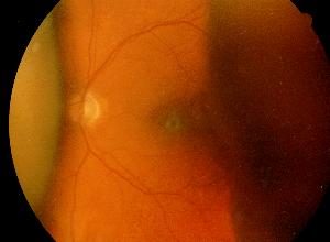
This 75 year old man had bilateral open angle glaucoma which were poorly controlled with topical medication. A left trabeculectomy was performed and he complained of reduced visual field 2 day post-operative. The above appearance was seen on fundoscopy.a. What does the picture show?
Choroidal detachment.
There is the characteristic brown-orange, solid appearing elevation with smooth, convex surface.
b. What are the possible causes of such appearances? What is the most likely cause in this patient?The following causes may cause choroidal detachment:c. How would you manage this patient?Choroidal detachment which occurs post-trabeculectomy is most likely to be caused by ocular hypotony. The intraocular pressure is usually less than 6 mmHg.
- Ocular hypotony
wound leak
glaucoma filter
penetrating ocular trauma- Elevated uveal venous pressure
arteriovenous fistula
vortex vein compression as by scleral buckle- Malignant hypertension
- Inflammatory processes
photocoagulation
uveitis
posterior scleritis- Neoplastic conditions
metastatic carcinoma
malignant melanomaThe key examination is to look for excessive aqueous drainage such as wound leak. Otherwise, the detachment does not normally need drainage as it usually resolves when the intraocuar pressure raised above 8 mmHg.
| Click here for the questions | Click here for the main page | Click here for FRCOphth/MRCOphth
/FRCS tutorials |