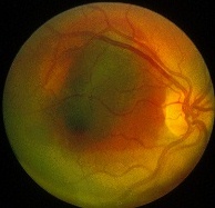
Subretinal, sub-RPE and choroidal haemorrhages
(Figure on the left shows the presence of all three haemorrhages
in a patient with traumatic choroidal rupture)All these haemorrhages have the same differential diagnosis and
they often co-exist. In the case of subretinal haemorrhage, the
haemorrhage is bright red and large with indistinct outline. In
sub-RPE haemorrhage, the blood appears dark-red and have
well-defined outline. In choroidal haemorrhage, the blood is
also dark-red but unlike sub-RPE haemorrhage, it is often extensive.
- bacterial endocarditis
- leukaemia
- severe anaemia
- HIV retinopathy
- multiple myeloma
Return to the top
- diabetes
- retinal vein occlusion
- retinal artery macroaneurysm
- malignant hypertension
- Coat's disease
- choroidal neovascularization
- radiation retinopathy
- retinal telangiectasia
Return to the top
- Pre-proliferative diabetic retinopathy
- retinal vein occlusion
- branch retinal artery occlusion
- hypertension
- HIV retinopathy
- ocular ischaemic syndrome
- auto-immune disorders
- SLE
- scleroderma
- Purtscher retinopathy
- haematological disorders
Return to the top
- diabetes mellitus
- old retinal vein occlusion
- retinal artery occlusion
- ocular ischaemia
ocular ischaemic syndrome
aortic arch syndrome (atherosclerotic disease, Takayasu's disease)
carotid-cavernous sinus fistula
- retinopathy of prematurity
- proliferative haemoglobinopathies
sickle cell haemoglobin C (SC)
sickle cell disease (SS)
sickle cell beta-thalassemia (S-thal)
- hyperviscosity syndrome
Waldenstrom's macroglobulinaemia
multiple myeloma
cryoglobulinaemia- Embolic disease
intravenous drug user
rheumatic heart disease
- Inflammatory disorder
sarcoidosis
Behcet's disease
par planitis
birdshot chorioretinopathy
acute retinal necrosis syndrome
vasculitis (both systemic and localized tot he retina)infectious causes
systemic lupus erythematosus
polyarteritis nodosa
Eale's disease
tuberculosis
Lyme disease
syphilis- Miscellaneous
radiation injury
familial exudative vitreoretinopathy
incontinentia pigmenti
long-standing retinal detachment
after encircling buckling procedures
primary systemic amyloidosis
hereditary haemorrhagic telangiectasia (Rendu-Osler-Weber disease)
Return to the top
- idiopathic
- infectious causes
herpes zoster virus
herpes simplex virus
cytomegalovirus
acquired immune deficiency syndrome (AIDS)
syphilis
tuberculosis
toxoplasmosis- Collagen vascular diseases
systemic lupus erythematosus
polyarteritis nodosa
Wegener's granulomatosis
giant cell arteritis
scleroderma- Other systemic diseases
sarcoidosis
Behcet's disease
multiple sclerosis
malignancy
ocular ischaemic syndrome
Crohn's disease
Churg-Strauss syndrome
Whipple's disease- Miscellaneous
Return to the top
acute frosted retinal periphlebitis
Eales' disease
pars planitis
- fibrinoplatelet from carotid diseases
- cholesterol emboli from carotid diseases
- calcific emboli from vulvular diseases
- infective embolic from bacterial endocarditis
- atrial myxoma
- fat emboli
- talc emboli
- metastatic tumour
- amniotic fluid
Return to the top
- systemic hypertension
- retinal artery occlusion
- retinitis pigmentosa
Venous dilatation and tortuosity:
- central retinal retinal vein occlusion
- preproliferative diabetic retinopathy
- hyperviscosity syndrome
- ocular ischaemic syndrome
- cartoid-cavernous fistula
- primary antiphospholipid antibody syndrome
- Fabry's disease
- mannosidosis
macular oedema (caused either by: 1. breakdown of the blood retina barrier
Return to the top
or 2. breakdown of the retinal pigment epithelium barrier)
white dot syndromes
- vascular causes
background diabetic retinopathy
retinal vein occlusion- secondary to other maculopathies
preretinal macular fibrosis
vitreomacular traction syndrome
age-related macular degeneration
- chronic intraocular inflammation
- retinal dystrophy
retinitis pigmentosa
gyrate atrophy- iatrogenic
cataract surgery
YAG capsulotomy
retinal detachment surgery
retinal cryotherapy
retinal laser photocoagulation
topical adrenaline eye drop in aphakia patients- others
Return to the top
high doses of nicotinic acid
familial dominant
Panuveitis
without inflammation
- retinitis punctata albescens
- fundus albipunctus
- fundus flavimaculatus
- Doyne honeycomb dystrophy
- malattia levantinese
- beign retinal syndrome
- Alport's disease
- Vitamin A deficiency
with inflammation
- multiple evanescent white syndrome
- acute posterior multifocal placoid pigment epitheliopathy
- punctate inner choroidopathy
- birdshot chorioretinopathy
- diffuse unilateral subacute neuroretinits
- acute retinal pigment epithelitis
- serpiginous choroiditis
atrophic maculopathies
- sarcoidosis
- Behcet's disease
- Vogt-Koyanagi-Harada's syndrome
- sympathetic ophthalmitis
- tuberculous choroiditis
- syphilis
bull's eye maculopathies
- dry age-related macular degeneration
- myopic maculopathy
- end-stage Stargardt's macular dystrophy
- central areolar choroidal dystrophy
- end-stage Best disease
- end-stage North Carolina macular dystrophy
choroidal neovascularization
- toxic drug exposure
chloroquine
hydoxychloroquine
clofazimine- retina dystrophy
cone dystrophy
Stargardt's disease
benign annular macular dystrophy
retinitis pigmentosa- systemic
neuronal ceroid lipofuscinosis
olivopontocerebellar atrophy
Hallervorden-Spatz syndrome
Bardet-Biedl syndrome
- age-related macular degeneration
- high myopia
- angioid streaks
- choroidal rupture and any pre-existing scars
- central serous retinopathy
- optic disc abnormalities
optic disc drusen
optic pits
coloboma- infectious
toxoplasmosis
toxocariasis
congenital rubella- inflammatory conditions
ocular histoplasmosis syndrome
sarcoidosis
Vogt-Koyanagi-Harada's disease
serpiginous choroiditis
acute posterior multifocal placoid pigment epitheliopathy- tumours
choroidal melanoma
choroidal naevus
choroidal haemangioma
choroidal osteoma- vascular causes
branch retinal vein occlusion
central retinal artery occlusion
parafoveal telangiectasia- retinal dystrophy
Best's disease
Stargardt's disease
adult foveal macular dystrophy
- idiopathic
- vascular causes
proliferative diabetic retinopathy
branch retinal vein occlusion- retinal hole or tear
- iatrogenic
pan-photocoagulation
vitreoretinal surgery- inflammatory eye diseases
true hole
- idiopathic
- traumatic
- high myopia
- chronic cystoid macular oedema
pseudo-hole
- epiretinal membrane
cherry red spots
- drug-induced
tamoxifen
canthaxanthin
talc
methoxyflurance- metabolic disorders
cystinosis
primary oxalosis type 1- others
Bietti retinal dystrophy
Sjogren-Larsson syndrome
isolated pigmented lesion
- central retinal artery occlusion
- Tay-Sachs disease
- Sandhoff disease
- Niemann-Pick disease
- generalized gangliosidosis
- sialidosis types I and II
isolated pale lesion
- choroidal naevus
- choroidal melanoma
- retinal pigment epithelium hamartoma
- congenital retinal pigment epithelium hypertrophy
Return to the top
- congenital retinal pigment epithelium atrophy
- amelanotic naevus
- amelanotic melanoma
- coloboma of the retina and choroid
- choroidal metastatic tumour
- retinal astrocytoma
- retinoblastoma
Return to the top
- multiple congenital hypertrophy of the retinal pigment epithelium
- rubella retinopathy
- congenital syphilis
- watermark in retinal detachment
- retinitis pigmentosa
- age-related peripheral retinal pigmentary changes
- female carriers of choroideremia
Return to the top
Return to the top
- congenital
extensive retinal nerve fiber myelination
coloboma of the retina and choroid- hereditary disorders
albinism
gyrate atrophy
choroideremia
diffuse choroidal atrophy- high myopia
- vascular
acute retinal ischaemia
- trauma
commotio retinae
Angioid streaks
- Idiopathic
- Ocular causes
hypotony
choroidal tumour
scleral buckling- Orbital causes
Return to the top
thyroid ophthalmopathy
pseudotumour
other orbital tumour
Retinal detachments
- Idiopathic
- Skin disorders
pseudoxanthoma elasticum
Ehlers-Danlos' syndrome- Bone
Paget disease
- Endocrine disorder
acromegaly
- Blood
Return to the top
sickle cell anaemia
abetalipoproteinaemia
thalassemia
hereditary spherocytosis
Choroidal detachments
- rhegamatogenous retinal detachment
- tractional retinal detachment
proliferative retinopathies
penetrating ocular trauma
severe intraocular inflammation- exudative retinal detachment
Return to the top
choroidal tumours
melanoma
intraocular inflammation
other primary tumour
metastatic tumourVogt-Koyanagi-Harada's disease
leaking subretinal vessels
posterior scleritischoroidal neovascularisation
systemic condition
retinal telangiectasiatoxaemia of pregnancy
others
malignant hypertension
hypoproteinaemic states such as nephrotic syndromeretinal photocoagulation
uveal effusion syndrome
- ocular hypotony
wound leak
glaucoma filter
cyclodialysis cleft
penetrating ocular trauma- elevated veal venous pressure
arteriovenous fistula
Sturge-Weber syndrome
vortex vein compression by scleral buckle- malignant hypertension
- inflammatory factors
after photocoagulation or cryotherapy
scleritis
uveitis- neoplastic conditions
metastatic carcinoma
malignant melanoma
lymphoid, leukemic or melanocytic choroidal infiltration- secondary to abnormal sclera
Return to the top
nanophthalmos
idiopathic uveal effusion syndrome