The duty of the referring doctor is to differentiate conditions which
do not require urgent ocular referrals from those which can be potentially
sight-threatening.
The most common causes of swollen eyelids are:
Chalazion
The eyelids contain many different glands which can become
blocked and superinfected. The resulting condition is termed hordeolum.
Presentation:
-
redness, swelling, and pain in the eyelid
-
they may be associated conjunctivitis and purulent discharge
Examination:
-
The visual acuity is normal unless the swelling is big and
right in the centre of the upper lid which can distort vision through its
mass effect on the cornea.
-
The swelling may be at the base of an eyelash (sty
or external hordeolum) or deep within the lid (meibomianitis or internal
hordeolum)
Management:
-
Topical antibiotic such as chloramphenicol drop is instilled
into the lower conjunctival sac four times a day.
-
If the lid swelling is extensive and severe, consider superimposed
orbital cellulitis which require systemic antibiotics
-
Referrals are not neccessary as the conditions resolves within
a few days.
-
Referred to the minor operating list if the swelling fails
to resolve after one week. This is mostly seen in internal hordeolum in
which a granuloma (chalazion) had developed. The treatment is incision
and curretage.
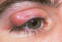 |
Figure 1.
This patient has a right upper lid chalazion. Note the
localized nature of the
swelling. This can be treated with topical chloramphenicol
by the GP. If the
swelling fails to resolve, refer the patient to the minor
operating list for
incision and curettage |
|
Orbital
cellulitis
This is a potentially sight-threatening condition and
the patient should be referred to the ophthlamologists for further management.
Sight loss may result from central retinal artery occlusion or optic nerve
inflammation. In adults the most common infection are Staphylococcus
aureus, Streptoccocus pyogenes or Streptoccus penumoniae. In children
, it is often secondary to infection in the adjacent sinuses and Haemophilia
influzae is an important pathogen.
Presentation:
-
Severe pain
-
Tense and red orbit with lid closure
-
Pyrexia
Examination
-
Intense swelling of the lids
-
Proptosis
-
Congestion of the conjunctival and episcleral vessels
-
Chemosis (swollen conjunctiva)
-
Double vision may occur due to poor eye movement in
a congested orbit.
Treatment:
-
Refer to the ophthalmologist within 24 hours.
-
Treatment require systemic antibiotics and analgesia.
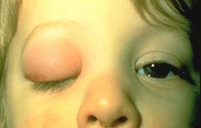 |
Figure 1.
This child has a typical appearance of orbital cellulitis
with swollen and tense right
eyelid and difficulty in openin the eye. Treatment should
involve admission with
intravenous antibiotics. |
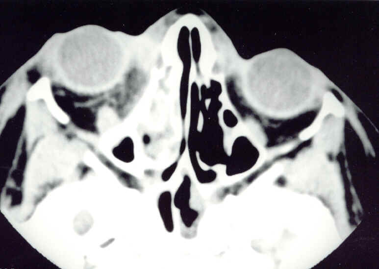 |
Figure 2.
This is the CT scan of a 7 year-old patient with a right
orbital cellulitis. Note the
presence of opacity in the right ethmoid sinus. The orbital
cellulitis is caused by
the spread of infection from ethmoid sinusitis. |
Return to the top
|
Herpes
zoster ophthalmicus

This is caused by reactivation of herpes zoster virus
in patient who previously had chickenpox. The eye is affected in 50% of
zoster ophthalmicus and is increased in patients with involvement of the
nasociliary nerve (rash at the tip of the nose).
Presentation:
-
pain in the distribution of the ophthalmic nerve followed
in a few days with vesicular eruption
Examination
-
Vesicular rash affecting the scalps and lids
-
Vision may be reduced with ocular involvement (keratitis
and anterior uveitis)
-
Swollen lids may make eye examination difficult
-
Ocular injections
-
Discharge from conjunctivitis
Management:
-
Oral acyclovir is useful in speeding up the resolution of
the rash
-
Analgesia should be given as the condition is very painful
-
Conjunctivitis is common and does not require treatment
-
Referred to the ophthalmologists within 24 hours from
seeing for exclusion of ocular involvement such as iritis and keratitis.
|
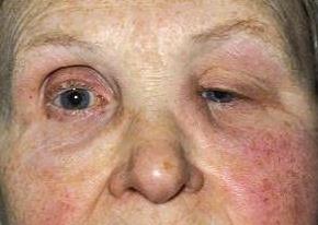
Figure 1.
This 78 year-old woman presented to the GP with a 3 day-history
of right sided headache.
The GP suspected giant cell arteritis but her ESR was
normal. Within 24 hours, she
developed this vesicular rash typical of herpes zoster
ophthlamicus. Note the distribution
of the rash which corresponds to the dermatome of the
ophthalmic nerve. She was referred
to the eye casualty and was found to have anterior uveitis.
She was treated with topical
steroid and mydriatic drops. |
Return to the top
|
Acute
dacryocystitis
This is caused by inflammation of the lacrimal sac. It
is often associated with obstruction of the nasolacrimal duct with watering
of the eye. Infection are often due to streptococcus and staphylococcus.
Presentation:
-
Painful swelling at the nasal side of the lower lid.
Examination:
-
Visual acuity is normal.
-
The swelling is tense and tender to touch
-
In severe cases, the whole of the lower lid may be swollen
due to superimposed cellulitis
Management:
-
Refer the patient to the ophthalmologists within 24 hours.
-
High dose systemic antibiotic is required either orally or
by intravenous.
-
Incision of the swelling should be avoided as this can cause
fistula formation
-
Most patient will require dacryocystorhninostomy (an artificial
passage is created between the lacrimal sac and the nasal cavity to bypass
the blockage) when the acute episode settle.
|
Figure 1.
This patient presented with a swellon and painful left
lower lid. Note the location of the
swelling which is diagnostic of acute dacryocystitis.
High dose oral antibiotics were given.
When the swelling settled, a dacryorhinostomy was performed
to prevent recurrence. |
|
|