The most common type of facial nerve palsy seen is
unilateral lower facial nerve palsy. Supranuclear facial nerve is
uncommon in ophthalmology examination because the
eyes are not involved. Remember not to use the term Bell's palsy
as synonymous with lower facial nerve palsy; Bell's
palsy is a diagnosis of exclusion when other known causes of lower
facial nerve palsy has been considered.
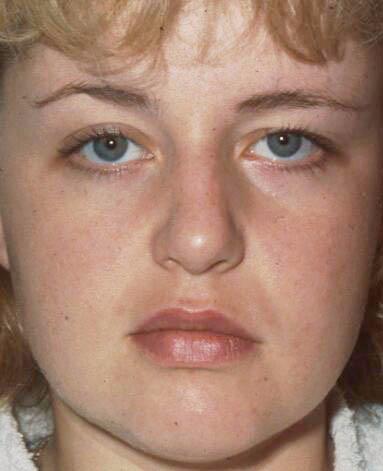
The patient has loss of nasolabial fold and the forehead
wrinkles of the affected side (in severe cases, there may be dropping
of the corner of the mouth and obvious ectropion). The
eyebrow of the affected side is lower (brow droop) and the upper lid
is retracted (ie. a wider palpebral aperture due to the
unopposed action of the levator). The blink rate on the affected side is
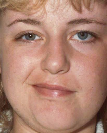
reduced. There is impaired blowing of the cheek, and
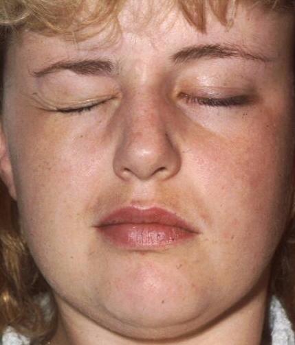
asymmetrical movement of the corner of the lip on smiling. On eyelid
closure, the affected side could not close the eye lid
fully (or it can be easily opened).
Important additional testing (to
assess the risk of exposure keratitis):
-
Check for Bell's phenomenon,
by observing if the eyes move up on attempted lid closure (you may need
to keep the
eyelids open to observe this but avoid hurting the patient)
-
Test the cornea sensation.
Further examination for any signs of aberrant regeneration
suggesting the lesion is long-standing, the following signs are
the most common:
-
Look for twitching of the mouth when the patient blink
-
Look for closure of the eye or asymmetrical narrowing of
the palpebral fissure on smiling
Further examination
for the cause of the facial nerve palsy
(the facial nerve is in close proximity to V and VI
nerve which may become involved if the lesion were intracranial):
-
Any signs of vesicles on the external
ear ? (Ramsey-Hunt's Syndrome)
-
Any signs of parotid swelling or scar
over the parotid gland? (sarcoidosis, parotid gland tumour or recent
parotid
gland operation)
-
Any signs of deafness? (previous mastoid abscess)
-
Any loss of cornea and facial sensation? (cerebellopontine
lesion)
-
Any scar behind the ear or behind the neck? (previous mastoid
operation or acoustic neuroma operation)
-
In patient with contralateral hemiplegia, test the eye movement
for ipsilateral gaze palsy and loss of facial sensation
from fifth nerve involvement (Foville's Syndrome).
|