| Differential Diagnosis
of Physical Signs in Cornea ferritin line
|
 |
| Differential Diagnosis
of Physical Signs in Cornea ferritin line
|
|
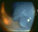
The cornea provides a variety of signs for the slit-lamp examination. These signs are easy to detect provided you examine each layer in turn and know how to use the various slit-lamp techniques. Usually more than one sign is present in the cornea. The following provide lists of differential diagnosis of common corneal signs seen in the examination.
Ferritin lines
(This seldom occurs in isolation in the examination. It is either a coincidental finding or associated with the corneal problem.)
- Hudson-Stahli's line (occurs in elderly and is found at the junction between the upper 2/3 and lower 1/3 of the cornea)
- Ferry's line (found anterior to a filtering bleb)
- Stocker's line ( found anterior to a pterygium)
- Fleischer's ring ( found at the base of keratoconus)
- foreign body
- other ( diseases which distort the surface of the cornea often causes iron deposition for example recurrent erosion syndrome, radial keratotomy)
Vortex keratopathy
(Also known as cornea verticillata; it is characterized by epithelial deposition which is arranged in a vortex fashion and usually begins just below the pupil and radiate outward. Like ferritin lines it is unlikely to be the only signs, if there is no other physical signs in the cornea ask to examine the posterior segment. For example, you may be given vortex keratopathy with tamoxifen deposition in the macula or vortex keratopathy with bull-eye maculopathy due to chloroquine toxicity.)
Corneal neovascularization
- Inherited
Fabry's disease
- Drug-induced
amiodarone
Return to the top
chloroquine/hydroxychloroquine
tamoxifen
chlorpomazine
indomethacin
(These may be superficial or deep in stroma. In the examination, the most common cases are phthisical eye, trauma, herpes simplex keratopathy, chemical burnt and ocular cicatricial pemphigoid.)
Band keratopathycontact lens wear trauma infections bacteria such as chlamydia, staphylococcus, pseudomonas
viruses such as herpes simplex and herpes zoster
protozoa such as onchocerca volvulus, leishmania brasiliensisalkali burns immunologic diseases Steven-Johnson's syndrome
graft rejection
cicatricial ocular pemphigoiddegenerative disorders pterygiumReturn to the top
Terrien's marginal degeneration
(It is found in the palpebral fissure. Although it may occur singly, in the examination you are likely to be given one with underlying ocular diseases for example chronic uveitis, interstitial keratic, phthsical eye or silicone oil in the anterior chamber)Ghost vessels
- Ocular causes
chronic inflammation (Still's disease and chronic uveitis)
interstitial keratitis
phthsical eye
silicone oil in the chamber- Systemic disease
hypercalcaemia
Return to the top
hyperparathyroidism
sarcoidosis
vitamin D intoxication
hypophosphataemia
Hereditary
dystrophic band keratopathy
(These usually occur in deep stroma in patients with interstitial keratitis. The chronic inflammation usually leaves a thin cornea and possibly anterior or posterior synechiae. Although interstitial keratitis classically occurs in congenital syphilis in which case the condition is invariably bilateral, it can also occur in a variety of infection. The most likely case in the examination is congenital syphilis and the patient may have the typical facial features of saddle nose, deafness and Hutchinson's teeth or may be normal. Avoid using words like syphilis (in front of the patient) when asked about the diagnosis or investigation instead used terms like St. Louis's disease or serology for Treponema pallidum.
The following is a differential diagnosis of interstitial keratitis:)
Breaks in Descemet's membrane
- Bacterial disease
congenital syphilis
tuberculosis
leprosy- Viral disease
herpes simple
herpes zoster
measles
mumps
rubella- Parasitic disease
leishmaniasis
onchocerciasis
cysticerosis- Unknown aetiology
Cogan's syndrome
Return to the top
(This is a common sign in the examination and the most common cause in the examination is bulphthalmos. Therefore, look out for differences in corneal size. )congenital glaucoma advanced keratoconus trauma (for example forcep delivery) Return to the top
Peripheral corneal thinning
(In the clinical examination, the most common cases will include rheumatoid arthritis, Mooren's ulcer and Terrein's marginal degeneration. However, in your differential diagnosis do not forget to mention more common condition such as bacterial infection or recurrent marginal keratitis.)Infectious Local immune diseases marginal keratitis
phylyctenulosis
vernal keratoconjunctivitis
Mooren's ulcer
superior limbic keratoconjunctivitisDegenerative disorders Terrien's marginal degenerationSystemic immune diseases Collagen vascular diseases
Return to the toprheumatoid arthritis
Dermatologic conditions
systemic lupus erythematosus
relapsing polychondritis
Wegener's granulomatosisrosacea
Inflammatory bowel diseasesCrohn's disease
Blood disorders
ulcerative colitisleukaemia
Corneal oedema
(Another common sign in the examination. The sign may be missed if subtle unless you look at the corneal thickness and the present of clear spaces in the stroma. You must look for the underlying cause. The most common case is pseudophakic bullous keratopathy.)
Pigments on the endothelium
- Increased intraocular pressure
congenital glaucoma
angle-closure glaucoma
neovascular glaucoma- Endothelium decompensation
inherited
congenital hereditary endothelial dystrophy
trauma
posterior polymorphous dystrophy
Fuch's dystrophy
post-operative
infectious
corneal ulcer
endopthalmitis
inflammationchronic uveitis
Return to the top
graft rejection
unknown
iridocorneal endothelial syndrome
(The typical case in the examination is Krukenberg's spindle in patient with pigment dispersion syndrome. However, pigments can also occur in various conditions and a detailed examination of the anterior segment may provide the clues.)
- pigment dispersion syndrome
- pseudoexfoliation syndrome
- post-cataract surgery
- previous corneal performation in which the iris was incarcerated in the wound leaving behing pigment
- trauma
- idiopathic
Return to the top
Keratic precipitates in the endothelium
(The most common case is Fuch's heterochormic cyclitis in which there are multiple discrete KP evenly distributed in the endothelium. Occasionally you may be given a patient with mutton fat KP and asked to give a differential diagnosis. Possible cases include sarcoidosis (look for nodules on the iris), toxoplasmosis (there may be chorioretinitis scar in the posterior segment) and rarely sympathetic ophthalmia (look for signs of ocular damage in the opposite eye)
The following list is for mutton fat KP ie. granulomatous inflammation.)
- Infectious
tuberculosis
syphilis
Lyme disease
toxoplasmosis
propiobacterium acnes- inflammation
sarcoidosis
Return to the top
sympathetic ophthalmia
phacoanaphylactic uveitis
Vogt-Koyanagi-Harada's syndrome