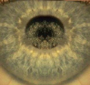
In the examination, the most common dystrophies you
are likely to see are Reis-Buckler's, lattice, granular,
macular and Fuch's endothelial dystrophies. As there
are many different types of dystrophies, you may well be
given less common types. Begin your presentation by
describing which layer (epithelium, anterior or posterior
stroma or endothelium) is involved, the pattern of
the opacities (crystalline, vesicles, lines or circular etc), and
if the opacities involves the centre or the periphery
of the cornea.
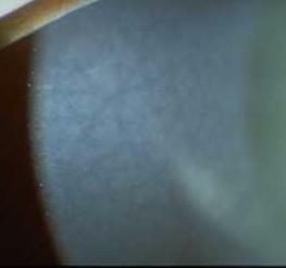
Schynyder central crystalline dystrophy
There are multiple small white crystals in the centre
of the cornea just below the Bowman's membranes.
The surface of the cornea is uninvolved and there may
be corneal arcus
Central cloudy dystrophy of Francõis
There are multiple greyish nebulous opacities separated
by crack like clear zones (the changes may resemble
crocodile shagreen). The opacities are denser in the
centre and the posterior aspect of the cornea.
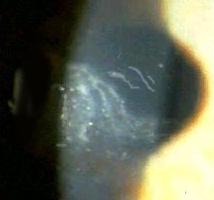
Posterior polymorphous dystrophy
There are multiple vesicles in the endothelium. There
are also multiple greyish opacities which may be curvilinear
or has scalloped edges. In some patients there may be
stromal oedema or abnormal iris shape such as corectopia
or iridocorneal adhesion (the changes may resemble ICE
syndrome but the condition is bilateral)
Map-dot-fingerprint dystrophy (Cogan microcystic dystrophy)
Although a common epithelial dystrophy, it seldom appears
in the examination.
There are multiple opacities in the epithelium and has
shapes resembling map lines, dots/microcysts or fingerprint
lines.
. |