1. The ultrasound relies on sound echo. What is an echo?
How can it be used to detect the length of a body
structures? What types of frequency
is used in medical ultrasound and why is this important?
2. How is the ultrasound used
in medicine produced?
3. How
is the ultrasound reflected by the body strucutres detected?
4. What is acoustic impendence? How does acoustic
impendence affect the ultrasound scan?
5. What does the A in A-scan stand for?
How is it produced?
6. What does the B in B-scan stand for? How is it produced?
7. What are the advantages and
disadvantages of ultrasound used in medicine?
8. What is real-time B
scan? What is doppler ultrasound?
1. The ultrasound relies
on sound echo. What is an echo? How can it be used to detect the length
of
a body structures? What
types of frequency is used in medical ultrasound and why is this important?
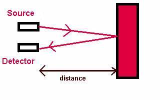
Whenever a sound wave moving in air hits a solid
surface, it reflects off it. This reflected sound is called an echo. The
same applies to a sound wave moving through water and hitting an obstacle.
If we know the speed of sound in the air or water, we
can calculate the distance to the obstacle. To do this we must measure
the time taken for a pulse of sound to travel to the object and back again:
The distance to the object and back is given
by
distance=speed x time
As this is the total distance that the sound
has travelled to the object and back, we must divide by 2 to find the one-way
distance.
This use of echoes is the basis of sonar (sound navigation
and ranging). The pulse of sound that is used should be short, and high
frequencies are usually used, as they travel further without being absorbed.
Sounds with a frequency above 20 kiloHertz (20 kHz) are called ultrasonic
(beyond the range of human hearing). The sounds used for sonar are well
into the ultrasonic range, with frequencies of 1 - 20 megaHertz (MHz).
In solving problems on sonar, remember that the speed
of sound itself varies from one material to another. The speed also depends
on temperature, pressure and other factors. Typical speeds are approximately
330 m/s in air, 1500 m/s in water and 5000 m/s in a metal.
2. How is the ultrasound
used in medicine produced?
The frequencies of ultrasound required for medical
imaging are in the range 1 - 20 MHz. These frequencies can be obtained
by using piezoelectric materials. When an electric field is placed across
a slice of one of these materials, the material contracts or expands. If
the electric field is reversed, the effect on the material is also reversed.
If the electric field keeps reversing, the crystal alternately contracts
and expands. So a rapidly alternating electric field causes the crystal
to vibrate.
The vibration is largest when the electric field stimulates
a natural frequency of the crystal - this is an example of resonance. The
vibrations are then passed through any adjacent materials, or into the
air as a longitudinal wave i.e. a sound wave is produced.
The piezoelectric effect occurs in a number of natural
crystals including quartz, but the most commonly used substance is a synthetic
ceramic, lead zirconate titanate. The crystal is cut into a slice with
a thickness equal to half a wavelength of the desired ultrasound frequency,
as this thickness ensures most of the energy is emitted at the fundamental
frequency.
3. How is the ultrasound reflected
by the body strucutres detected?
The piezoelectric effect also works in reverse.
If the crystal is squeezed or stretched, an electric field is produced
across it. So if ultrasound hits the crystal from outside, it will cause
the crystal to vibrate in and out, and this will produce an alternating
electric field. The resulting electrical signal can be amplified and processed
in a number of ways (see questions on A-scan and B-scan). So a second crystal
can be used to detect any returning ultrasound which has been reflected
from an obstacle.
Normally the transmitting and receiving crystals are built
into the same hand-held unit, which is a called an ultrasonic transducer
(generally, a transducer is any device to convert energy from one form
to another, usually to or from electrical energy.)
4. What is acoustic
impendence? How does acoustic impendence affect the ultrasound scan?
The exact fraction of the incident sound which is transmitted
or reflected depends on how different the two materials on each side of
the boundary are. This is described by the acoustic impedance of the materials,
which is related to the density of the material and the speed of sound
in the material. The greater the difference in impedance, the more sound
will be reflected rather than transmitted. Some typical impedances are
shown in the table below:
| Medium |
Impedance (in standard unit) |
| air |
0.000429 |
| water |
1.50 |
| blood |
1.59 |
| fat |
1.38 |
| muscle |
1.70 |
| bone |
6.50 |
Air and water have very different impedances, so that
a beam of ultrasound hitting a water surface is almost entirely reflected
away, and only a small amount enters the water. The same applies to a beam
trying to enter the eye from air.
Because of the impedance difference between air and skin,
a coupling medium helps to match the impedance of the crystal in the probe
more closely to the the impedance of the skin of the patient. The most
common coupling medium is a film of oil smeared on the patient's skin.
The operator requires needs to ensure that the probe is kept in continuous
contact with the oil, preventing air bubbles coming between the probe and
skin.
On the other hand, different body layers such as fat,
muscle and many body organs, have very similar impedances, so that most
of the beam will pass from one layer into the next, and only a small fraction
is reflected. In practice, this is not a problem; in fact the imaging technique
relies on it. To obtain a reasonable image with good resolution of an interface
between two layers, around 1% of a beam must be reflected, leaving a substantial
portion to continue on to further reflections.
5. What does the A
in A-scan stand for? How is it produced?
Each layer of tissue in the body produces a separate
reflection of the ultrasound signal. In the original type of scanner, these
reflections were simply displayed on the screen of a cathode ray oscilloscope.
This results in a
trace just like the Sonar trace below, where the x-axis
represents time, and the y-axis represents the
amplitude of the reflection i.e. how strongly the sound
is reflected.
Each layer producing a reflection shows up as a peak on
the trace. This gives rise to the name of the technique, an amplitude-scan
or A-scan. The diagram below shows an A-scan which could result from
the layers in the eye (this should be familiar to all ophthalmologist ever
performs biometry). The amplitude of the peaks depends on the difference
in acoustic impedance between the tissues on each side of each boundary.
It also depends on how much of the sound is absorbed as it travels through
each layer, an effect which complicates the interpretation of the scan.
 |
a = cornea spike
b = anterior lens spike
c = posterior lens spike
d = retinal spike
e = orbital spike |
6. What does the B in
B-scan stand for? How is it produced?
In this method the amplitude of each returning
signal is not simply displayed on a graph or CRO screen. Instead the amplitude
controls the brightness of the spot which represents this reflection, the
b for brightness giving rise to the name B-scan. So a single pulse of ultrasound
passing into a series of tissues will give rise to a series of spots, with
the brightness of the spots corresponding to the amplitude of the reflection
from different layers.
The largest amplitude gives rise to a spot with the greatest
brightness, here shown almost white. The smallest amplitude gives rise
to a spot which is almost black. Intermediate amplitudes give various shades
of gray. The area that does not give rise to any spike for example the
aqueous and the vitreous will appear black
 |
The corneal spike and the retinal spike
have the biggest amplitude and therefore appears nearly white. The posterior
lens spike has a lower amplitude than the anterior lens spike and therefore
appears darker. The aqueous and the vitreous allow the sound to pass through
with little impendence and therefore appears black. The lens subtance offers
some resistance and therefore does not appears as black. |
7. What are the advantages
and disadvantages of ultrasound used in medicine?
Advantages compared with other techniques
1. Ultrasound examinations are non-invasive
i.e. they do not require the body to be opened up, or anything
to be inserted into the body. This
is a major advantage compared to fibre-optic endoscopy, for example,
which may involve much more patient
discomfort as the probe is inserted.
2. Ultrasound methods are relatively inexpensive,
quick and convenient, compared to techniques such as
X-rays or MRI scans. The equipment
can be made portable, and the images can be stored electronically.
3. No harmful effects have been detected,
at the intensity levels used for examinations and imaging. This
contrasts with methods based on X-rays
or on radioactive isotopes, which have known risks associated
with them, and ultrasound methods
are preferred whenever possible. This is particularly relevant to
examination of expectant mothers.
4. Ultrasound is particularly suited to imaging
soft tissues such as the eye, heart and other internal organs,
and examining blood vessels.
Disadvantages of ultrasound compared with
other techniques
1. The major disadvantage is that the resolution
of images is often limited. This is being overcome as time
passes, but there are still many situations
where X-rays produce a much higher resolution.
2. Ultrasound is reflected very strongly on passing
from tissue to gas, or vice versa. This means that
ultrasound cannot be used for examinations
of areas of the body containing gas, such as the lung and the
digestive system.
3. Ultrasound also does not pass well through bone,
so that the method is of limited use in diagnosing
fractures. It is possible to obtain
quite good ultrasound scans of the brain, but much greater detail
is obtained by an MRI scan.
8. What is real-time
B scan? What is doppler ultrasound?
Real-time B-scans
Real-time B-scans allow body structures which are moving
to be investigated. The simplest type of scanner is just a speeded up version
of the 2-D B-scan , allowing a rapid series of still pictures to be built
up into a video of the movement. More sophisticated systems have an array
of transducers rather than just one pair of transmitter and detector, and
the current image is combined by computer with a recently stored image
to improve the overall quality.
Doppler Methods
When ultrasound is reflected from a moving surface, the
frequency of the sound is altered slightly in a manner that depends on
the speed of movement of the surface. This is due to the Doppler effect.
The Doppler effect is now commonly used in ultrasound
imaging to examine the movement of liquids, such as blood flow inarteries
and veins, allowing the location of blockages to be determine precisely.
Another common medical application is in foetal heart monitoring.
|