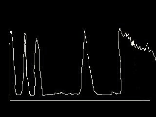
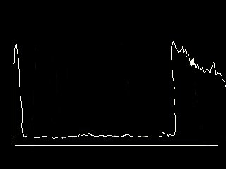
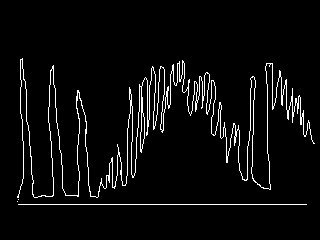
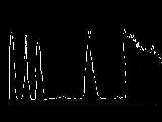
Common A scan (Amplitude scan) Cases in OSCE
(like all imagings, ultrasound scans should supplement clinical findings and 
 not be used alone in clinical diagnosis or management)

1. Most cases of A scan appearing in the OSCE are related to the biometry calculation.
    Common questions being:
    • the structures represented by each peaks (see diagram below)
a = cornea spike 
b = anterior lens spike 
c = posterior lens spike 
d = retinal spike 
e = orbital spike
    • the effect of silicone oil on the axial length (the value will be longer than expected as ultrasound travels slower in the silicone oil than vitreous)
    • the accuracy of the axial length(s)  ie, if the ultrasound scan is accurate as demonstrated by one retinal peak rather than several peaks; if there is significant differences between the two eyes. Note: the Guideline on Cataract Operation from the Royal College of Ophthalmologists finds that :
        - 96% of axial lengths fall within the range 21.0 to 25.5 mm and for 60% 
           this is between 22.5 and 24.5 mm
        - in the absence of pathology that might affect eye size (eg, unilateral 
          refractive error, coloboma or staphyloma), most individuals have similar 
          axial lengths in each eye
        .
        .
    2. The following scans may appear in the future examination as the examiners look for new 
        questions. Using the principles of ultrasound can you diagnose or give a differential diagnosis
        for the following scans ? (click on the scans for questions and answers)

Return to Index Page