Cataract
 

Hypermature cataract. Note the
wrinkling of the anterior capsule,
the lens has liquefied and leaks out
of the capsule.

A morgagnian cataract. The cortex 
has turned into milky liquid and the 
nucleus is displaced inferiorly.

A rosette cataract. This is seen in 
blunt trauma. Look for other signs 
in the posterior segment such as
choroidal tear or retinal detachment.

A lamellar cataract. There are 
opacities at various levels of the 
fetal nucleus. It is the most common 
type of congenital cataract. 

A posterior subcapsular cataract. 
Causes include steroid use, retinitis 
pigmentosa, atopic dermatitis 
diabetes and chronic uveitis.

A droplet cataract seen in a patient
with galactosaemia. 

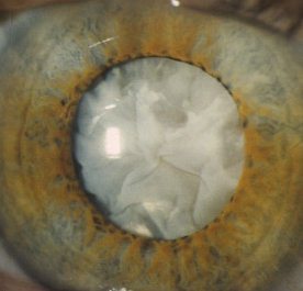
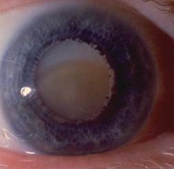
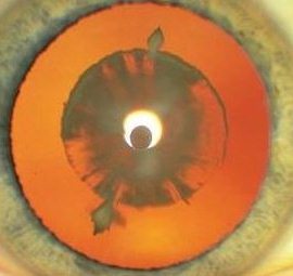
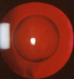
Cataract may be cortical, nuclear, subcapsular or in any combination. Examine the cataract with different forms of
biomicroscopic illumination so that you can describe the location of the cataract. For example, retroillumination is best
for anterior and posterior subcapsular opacities whereas direct focal slit illumination is best for examining the different
zones of the lens and thus locate the opacities. The location of the cataract can suggest the cause. 

In the examination:

    a. if the patient is young and has bilateral cataract. Consider the following:
    • atopic dermatitis (observe the face for dermatitis)
    • diabetic mellitus (examine the fundus for diabetic retinopathy
    • retinitis pigmentosa (examine the fundi for pigmentary changes)
    • myotonic dystrophy (note the typical facies of frontal balding, bilateral ptosis and delayed muscle

    • relaxation)
    b. in unilateral cataract. Look for:
    • Fuch's heterochromic uveitis
    • trauma
    • chronic uveitis
    • retinal detachment

Questions:

1. What happen to the lens in poorly controlled diabetes mellitus?

2. In which form of cataract would the patient gain a second sight and why?

3. How can cataract cause glaucoma?


Return to the main page