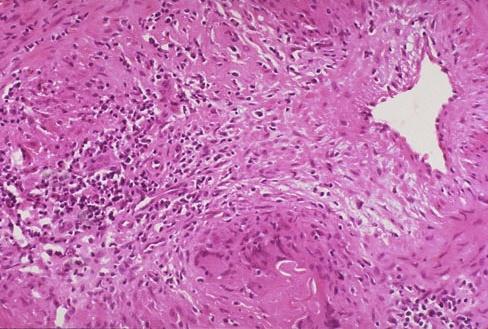
This is the histopathology of an artery taken from a patient with sudden visual loss. a. What does the picture show? b. What blood tests may be positive? c. What ocular signs may be present? Click here for the answers
a. What does the picture show?
b. What blood tests may be positive?
c. What ocular signs may be present?
Click here for the answers
This is the histopathology of an artery taken from a patient with sudden visual loss. a. What does the picture show? The histology shows focal granulomatous inflammation and narrowing of the arterial lumen. This is typical of giant cell arteritis. b. What blood tests may be positive? The ESR and C-reactive proteins are typically raised. There may be normochromic normocytic anaemia. There is usually a thrombocytaemia. c. What ocular signs may be present? Ischaemic optic neuropathy resulting from posterior ciliary artery occlusion Central retinal artery occlusion Amaurosis fugax Cortical blindness Anterior segment ischaemia. Click here for more questions
The histology shows focal granulomatous inflammation and narrowing of the arterial lumen. This is typical of giant cell arteritis.
Click here for more questions