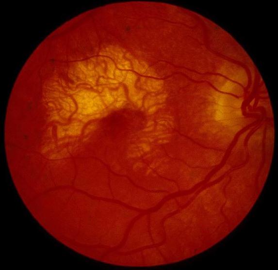
Figure 1 |
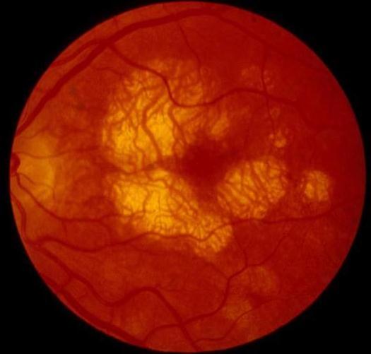
Figure 2 |
Medical Retina: Case fourSpecial thanks to Mr. Paul Parker, Chief Photographer of the Eye Hospital, Radcliffe Infirmary, Woodstock Road, Oxford for taking these pictures
|
Figure 1 |
Figure 2 |
This 48 year old woman has a ten year history of deteriorating vision in both eyes. Her visual acuity was 6/9 in the right eye and 6/18 in the left. Her mother and one of her brothers also suffer from poor vision from the age of 40. The above pictures were taken in one of her clinic visits.
a. What is the most likely diagnosis?
Central areolar choroidal atrophy.It is a bilateral symmetrical progressive dystrophy.
The early stage of the disease is non-specific with granular hyperpigmentation. The above pictures show the late stages of the disease. There are well demarcated area of retinal pigment epithelium atrophy with underlying loss of choriocapillaries leaving behind visible middle and large choroidal vessels.
b. How is this condition transmitted?The condition is an autosomal dominant disease affecting the macula. The abnormal gene is located in chromosome 17.c. What would you expect to find in the electrophysiology tests?The ERG and EOG are usually normal.d. What is the visual prognosis for patients with this condition?Typically the patients present in the third to fourth decade with symptom of central visual disturbance. The vision decreases as the atrophy progresses resulting in central scotoma by the 6th or 7th decade of life.
| Click here for questions | Click here for the main page | Click her for FRCOphth/MRCOphth
/FRCS tutorials |